[DBS seminar] Miroslav Vořechovský, "Model for Mechanistic Coupling of Patterning and Proliferation in Organoids by a 3D Vertex Model"
→
Europe/Ljubljana
Seminar room of physics (106) (IJS)
Seminar room of physics (106)
IJS
Jamova 39, 1000 Ljubljana, Slovenia
Description
A three-dimensional computational model inspired by the work Okuda and his collaborators [1], is developed to reveal the interaction between patterning and proliferation in epithelial tissue. The geometrical representation is based on Voronoi cells representing individual cells and differentiating the lateral, apical an basal cell faces. The viscous-damped system of forces controlling the shape of the multicellular tissue is derived from a potential featuring both volumetric and surface terms. Equipped with the cell rearrangements via cell-neighbor exchanges, the model allows for cell division and splitting while following the cell cycles.
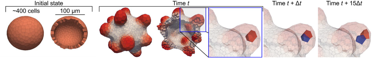
Fig. 1 Cell division during pattern-driven growth from initial single-layered spherical organoid.
Pattering is controlled by the interplay of two morphogens (activator and inhibitor) in a reaction-diffusion system [2]. The formation of patterns controls the cell proliferation in which the local concentration of activator which plays a role of a mitogen. The cell growth rate is modelled via Hill equation in which the exponent plays a central role in the presented coupling between patterning and proliferation.
The developed model is used to decipher the diverse roles of ERK and Akt signaling in organoid morphogenesis by matching the laboratory data. In our computational model of multicellular organoid growth, we have identified a feedback loop between the formation of patterns, influencing the rate of cell proliferation and consequently the tissue mass increase. This feedback loop critically impacts the subsequent patterning through a diffusive-reaction system of morphogens. The interplay between proliferation and patterning accurately captures experimentally observed variations in the shapes of growing mammary gland organoids in the presence of two types of FGF: WT and Stab. The integration of the model with laboratory data and its biological interpretation are presented in a complementary contribution.
REFERENCES
[1] Okuda, S., Inoue, Y. & Adachi, T. Three-dimensional vertex model for simulating multicellular morphogenesis. Biophysics and Physicobiology 12, (2015) 13–20. doi:10.2142/biophysico.12.0_13
[2] Gierer, A. & Meinhardt, H. A theory of biological pattern formation. Kybernetik 12, (1972) 30–39. doi:10.1007/bf00289234
Acknowledgement: Czech Science Foundation project no. GA23-04974S.